

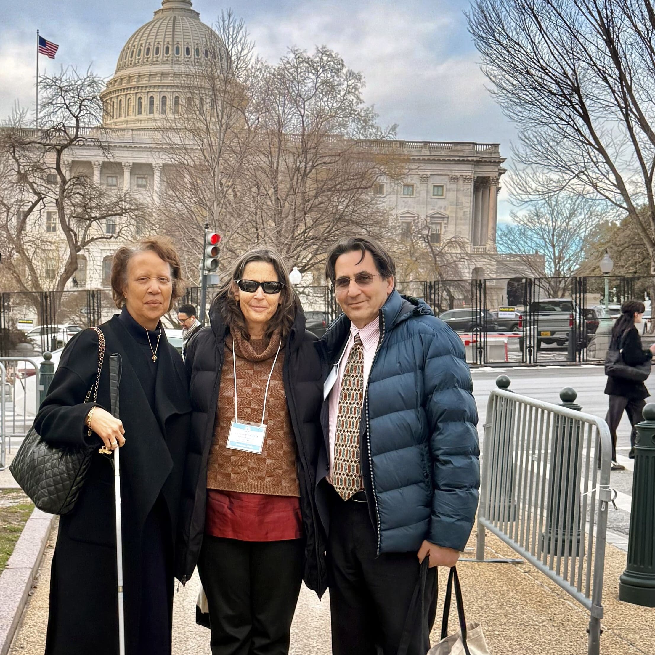
On February 24, 2026, AMDF staff joined with the National Alliance for Eye and Vision Research (NAEVR), and people living with macular degeneration to walk the halls of Congress. We sat across from legislators and their staff, shared what it’s like to lose your central vision, and made the case for why federal action is needed to help the over 20 million Americans living with some form of macular degeneration.
Why We Were There
We don’t need to tell you about the real-world impacts of living with macular degeneration. You’re already living it. But we do need to tell members of Congress. Macular degeneration and vision loss are not topics that are well understood by policy makers. So we need to raise our voices and concerns more.
Because even though macular degeneration is the leading cause of vision loss in Americans over 50, federal funding for eye disease research is not keeping pace leaving promising research projects on the table.
Even though central vision loss robs people of independence, Medicare still excludes coverage for the assistive devices that could help restore independence, and it’s the only disability for which this is the case.
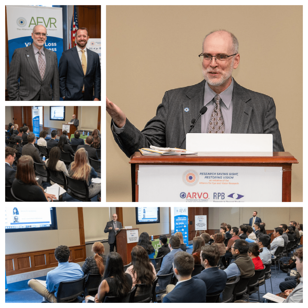
There’s more. AMDF’s Matthew Levine spoke eloquently on these topics at the Congressional Briefing, and you can watch that section below.
The people making decisions about these policies do so whether or not they’ve ever heard from someone living with AMD. Our job is to make sure they have.
In 2026, we are asking Congress to:
Fund the National Eye Institute for Fiscal Year 2027 at $1 Billion, to match growing public health needs. We urge Congressional support to increase this funding in the FY27 appropriations requests.
Reject any future proposed consolidation of the National Eye Institute (NEI) by maintaining the NEI as a dedicated institute within NIH in FY27 appropriations.
Support access to low vision assistive devices and services by urging the Centers for Medicare and Medicaid Services (CMS) to rescind the Medicare “Low Vision Aid Exclusion” at 42 C.F.R. § 411.15(b).
Protect Vision and Eye Health Funding in FY 2027 appropriations by restoring the Vision Health Initiative at the CDC to $6.5M to allow them to complete their critical work in community-level eye health interventions, surveillance, and research.
Join the bipartisan Congressional Vision Caucus, whose members are dedicated to strengthening and stimulating a national dialogue around policies related to vision loss, blindness, and visual impairments or disabilities.
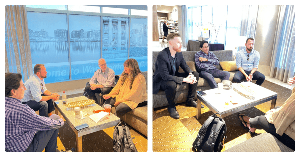
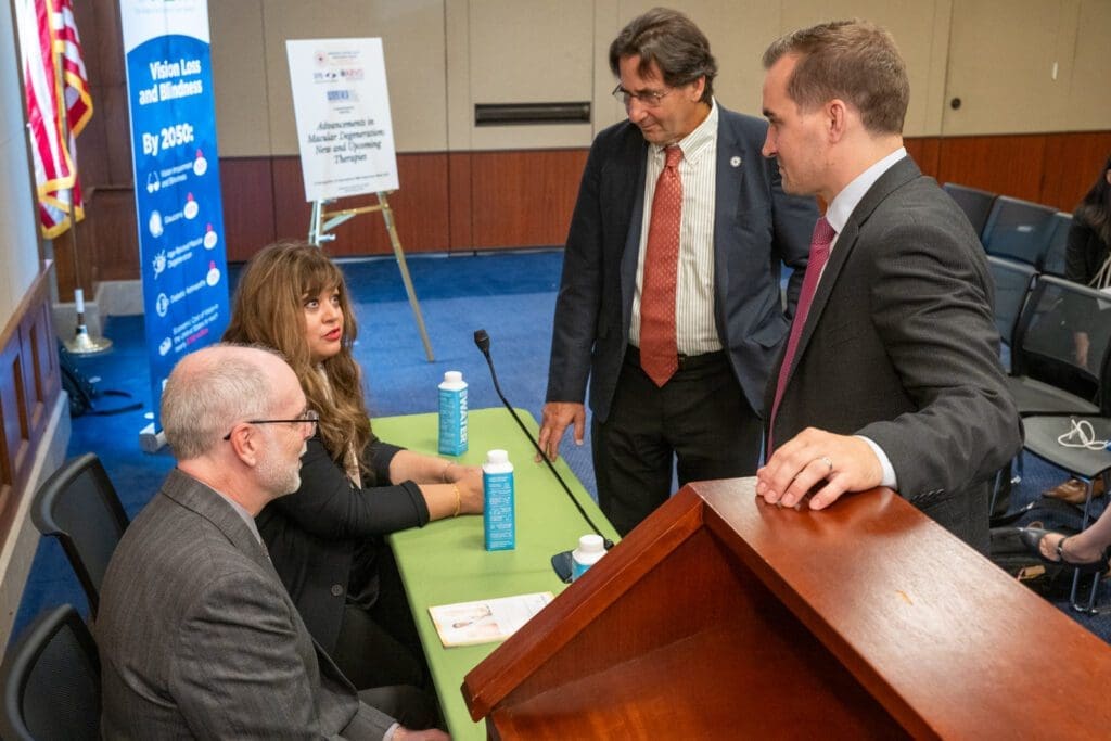
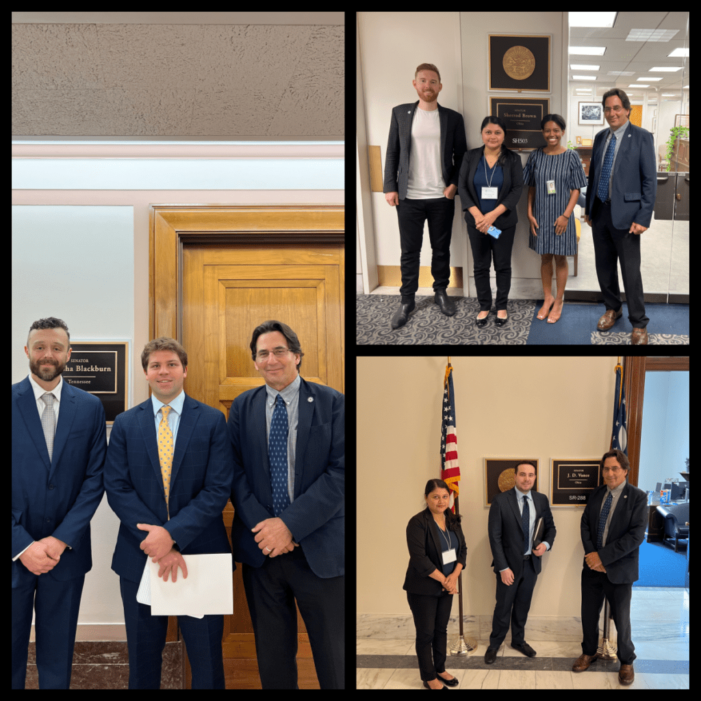
Congressional Office Visits
In addition to AMDF’s Matthew Levine, we were joined in our Congressional office visits by Dan Ignaszewski and Mikyla Bethune from NAEVR. Sadly, a snow storm prevented our founder, Chip Goehring, who was planning to attend along with AMDF Secretary Paul Gariepy, from driving to DC as planned.
We were also joined by AMDF Patient Advocate, Connie Hills, and patient advocate, Janice Samuel, from the Prevention of Blindness Society of Metropolitan Washington.
Together, this group had eight in-person meetings with the following offices:
- Sydney Lamb of Senator Richard Blumenthal’s office (CT)
- Karina Bravo of Senator Markey’s office (MA)
- Maisie Ruddy of Congressman Glenn Ivey’s office (MD)
- Chelsey Rice-Davis of Senator Chris Murphy’s office (CT)
- Hanna Vohra of Senator Chris Van Hollen’s office (MD)
- Emmett Jamieson of Senator Eric Schmitt’s office (MO)
- Thomas Greco of Senator Alex Padilla’s office (CA)
- David Seyferth of Congresswoman’s Jahanna Hayes’ office (CT)
We left the 2026 AMDF Congressional Report behind at each office. Click the image below to download the PDF.
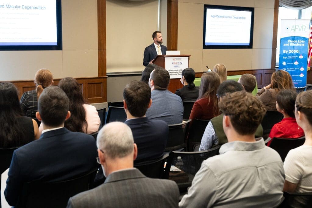
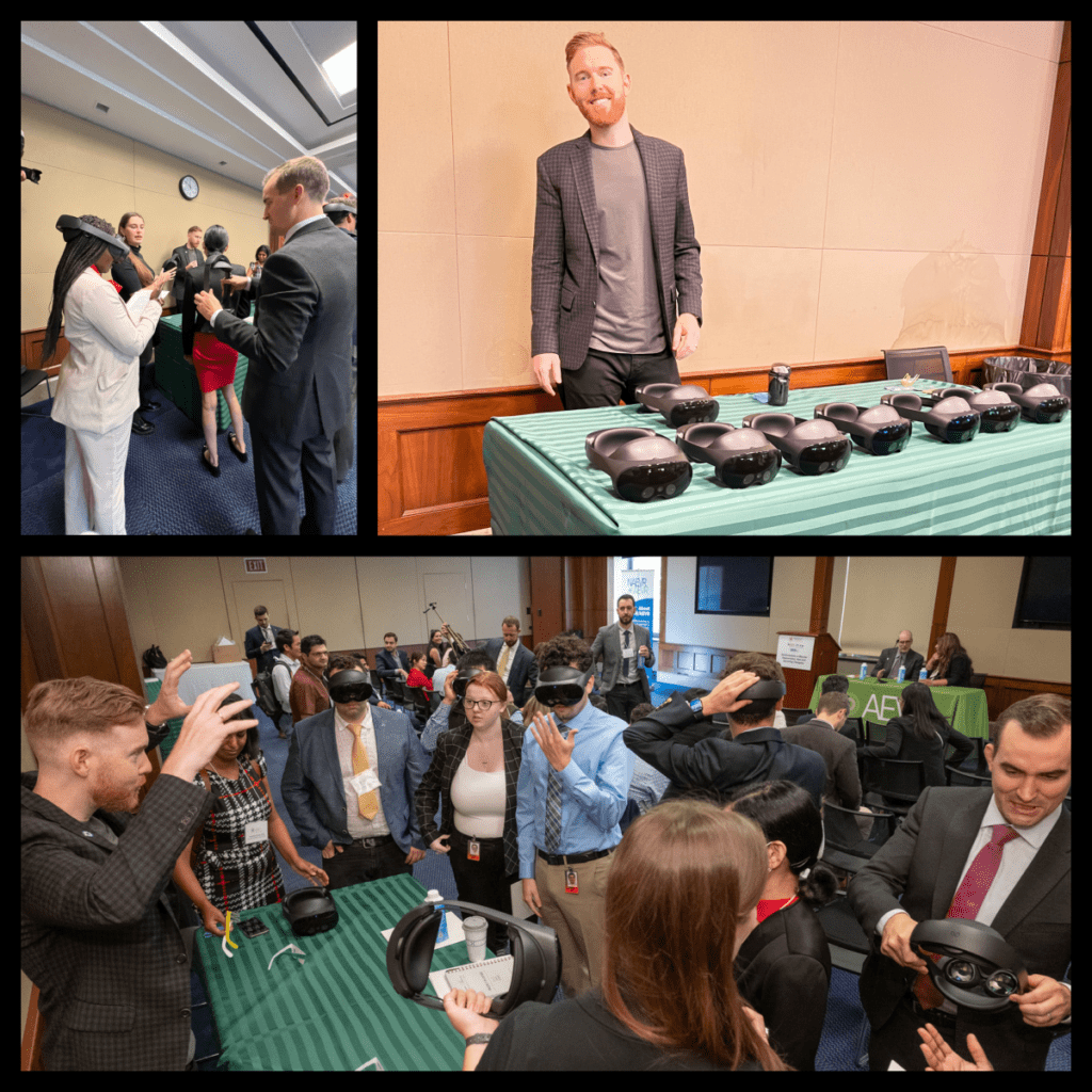
The Congressional Briefing
At noon, we co-hosted a Congressional Briefing with NAEVR. Invitations went out to all members of Congress to attend or send an appropriate staffer to learn about macular degeneration and what we’re asking.
The speaker lineup included AMDF and NEI funded scientist Dr. Rajendra Apte (who couldn’t make it due to the epic snowstorm, but sent a recording of his presentation); AMDF patient advocate Connie Hills; Diane Bovenkamp, Vice President of Scientific Affairs at Bright Focus Foundation; and Matthew Levine, Director of Grants, Advocacy, and Partnerships at AMDF.
You can view the Congressional Briefing in its entirety below:
In attendance at the briefing were:
- Riya Singh, Legislative Intern for Senator Michael Bennet (CO)
- Angel Osander, Intern for Senator Van Hollen (MD)
- Michelle Elele, Intern for Rep. Marc Veasey (TX)
- Kwasi Irving Jr, Intern for Rep. Kweisi Mfume (MD)
- Evasion Alston, Intern for Rep. Linda Sanchez (CA)
- Ashley Ordering, Intern for Rep. Joe Neguse (CO)
- Alvin Xu, Intern for Rep. Veronica Escobar (TX)
- Vanessa Baker, Legislative Aide for Rep. Lori Trahan (MA)
- Chris Beny, Scheduler for Janelle Bynum (OR)
- Peace Ade, Scheduler for Rep. LaMonica McIver (NJ)
- Gwen Che, Fellow for Senator Amy Klobuchar (MN)
- Alexander Gristina, Legislative Assistant for Congressman Frank Pallone Jr (NJ)
- Jordan, Intern (office not listed)
- A’Milliana McNeil, Associate at VHAC
- Jimmy Liu, Director of Vision Science Program at Bright Focus Foundation
- Rachel Gandell Tetlow, President of Government & Political Affairs, Prevent Blindness
- Lauren Chong and Isabella Moore, interns at the Federation of Associations for Brain & Behavior Sciences
What’s Next and What You Can Do
Check the lists above. If your representative isn’t listed, contact them. Ask them what they’re doing to protect constituents like you living with macular degeneration. Point them to the Congressional Briefing recording and the 2026 AMDF Congressional Report.
And if you DO see your congressman/woman’s name on the list above, thank them for taking a meeting with us, or for sending someone to the Congressional Briefing. It lets them know this community is paying attention.
If you would like call scripts, or a form letter, you can find these on our Access in Sight page.