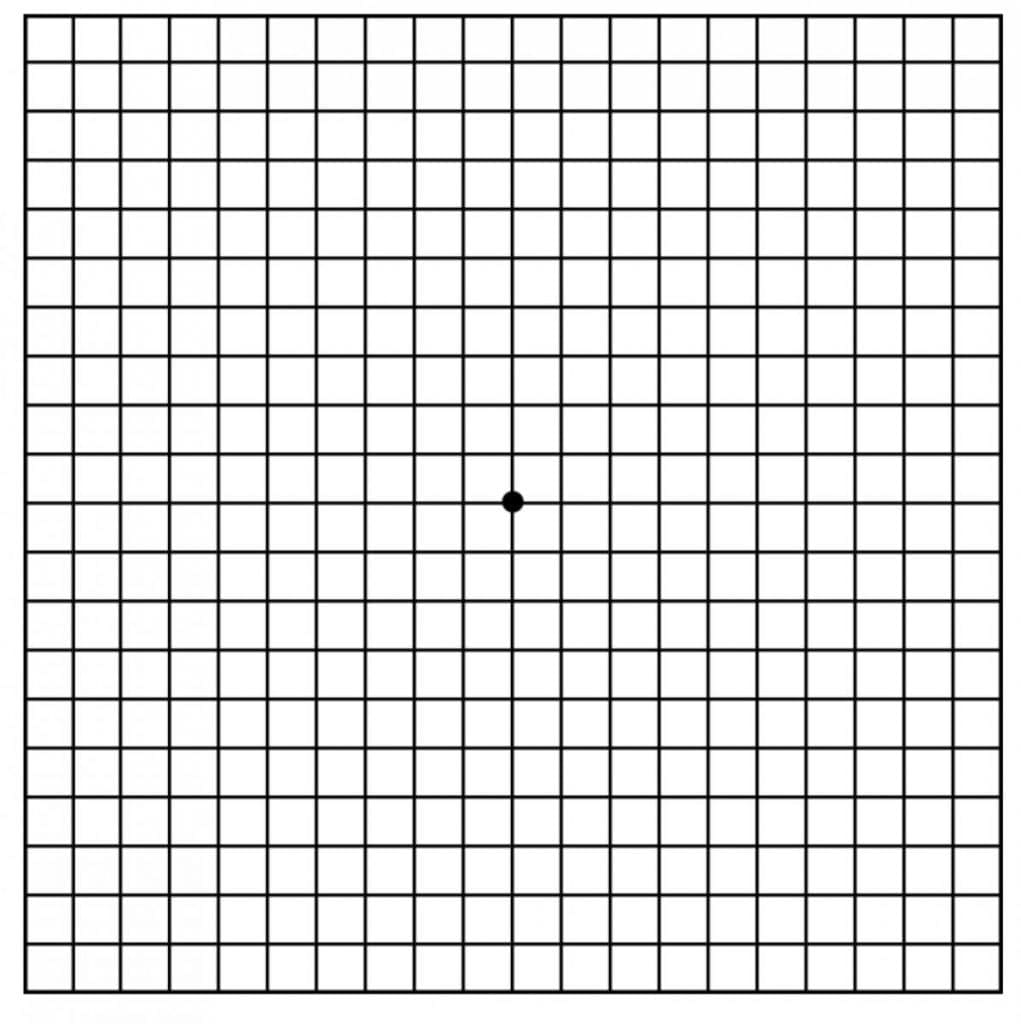
The first signs of Age-related Macular Degeneration are typically discovered by an eye doctor in an annual dilated eye exam. They include the presence of drusen – tiny but visible heaps of cell waste on the surface of the retina – and pigment changes in the macula. Often these signs of AMD are present long before any changes are noticeable in a person’s vision. In fact, nearly everyone over age 50 has at least one small drusen. Standard screening tests include the visual acuity exam (the letter chart with an E at the top) and an Amsler grid, which looks like graph paper.

Click here for a larger Amsler grid.
The Amsler grid is used to check whether lines look wavy or distorted, or whether areas of the visual field are missing. Most of the advanced diagnostics for studying the presence or progression of macular degeneration involve making images of the fundus (the inside back of the eyeball) and the retina. The technology is constantly being updated, but currently includes:
Fundus Fluorescein Angiography (FFA)
A yellow dye called fluorescein is injected into a vein in the arm or hand. Sequential photographs are taken of the eye, using a camera that sits on a table (the person sits in front of it). The images show blood flow and possible leakage (wet AMD) in the retina and choroid. This method of imaging takes about 20 minutes altogether and requires dilation drops. Urine and saliva are sometimes dark orange afterwards, a result of the body metabolizing the dye.
Optical Coherence Tomography (OCT)
In this common, non-invasive (no injections) imaging method, infrared light waves are used to make cross-section photographs of the retina and choroid layer. OCT provides extremely useful information about drusen, retinal structure, new blood vessels, and hemorrhaging. The person gets dilation drops then sits in front of the camera with his or her chin in a chin rest. The imaging equipment takes about 15 minutes.
Fundus Autofluorescence Imaging (AF)
This is an imaging method – also non-invasive – which uses the body’s natural fluorescence to study the retina. Certain structures (fluorophores) in the body will light up when exposed to certain wavelengths of light. These include retinal pigment epithelial cells and lipofuscin (the residue of RPE waste). This imaging method is often used to study late dry AMD, or geographic atrophy because atrophied sections of the eye do not light up.
Genetic Testing
Another diagnostic for young onset macular degeneration is genetic testing, currently available for Stargardt disease and other juvenile macular degeneration diseases. Genetic testing would indicate the presence of the recessive gene which causes Stargardt disease. At this point in time, genetic testing for Stargardt serves only two purposes: the first is for family planning; the second is to diagnose Stargardt in young people with symptoms of the disease. But researchers are working at the level of the human genome, the code that tells the body how to build and rebuild itself. They believe that, in the next few years, gene therapy will lead to the replacement of defective genes within the very DNA of a person with Stargardt or AMD. The American Macular Degeneration Foundation supports this research.

