


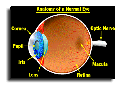
Cornea
The transparent “front window” of the eye. It is a thick, nearly circular structure covering the lens. The cornea is an important part of the focusing system of the eye.
Pupil
The round black hole in the center of the iris. The size of the pupil changes automatically to control the amount of light entering the eye.
Iris
The pigmented (colored) membrane of the eye, the iris is located between the cornea and the lens. Its color varies from pale blue to dark brown.
Lens
A transparent biconvex structure located behind the iris. It focuses light rays entering through the pupil in order to form an image on the retina.
Retina
A thin multi-layered membrane which lines the inside back two-thirds of the eye. It is composed of millions of visual cells and it is connected by the optic nerve to the brain. The retina receives light and sends electrical impulses to the brain that result in sight. The light sensitive retina consists of four major layers: the outer neural layer, containing nerve cells and blood vessels; the photoreceptor layer, a single layer that contains the light sensing rods and cones; the retinal pigment epithelium (RPE) and the choroid, consisting of connective tissue and capillaries.
Macula
An area of the eye near the center of the retina where visual perception is most acute. The macula is responsible for the sharp, straight-ahead vision that is used for seeing fine detail, reading, driving, and recognizing faces. It is one hundred times more sensitive to detail than the peripheral retina. The macula is sometimes referred to as “the bull’s eye center of the retina.”
Optic Nerve
Cable-like structure composed of thousands of nerve fibers that connect the macula and retina with the brain. The optic nerve carries electrical impulses from the macula and retina to the processing center of the brain where they are interpreted into clear, colorful sight.